
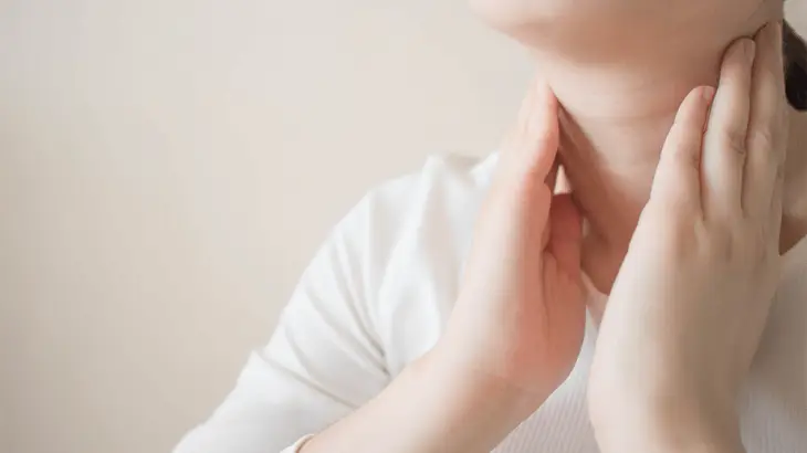
Goiter (Enlargement of the Thyroid Gland)
The thyroid gland is located in the front of the neck, below the cartilage, and consists of two parts, the right and left lobes. It secretes the hormones thyroxine (T4) and triiodothyronine (T3).
The thyroid gland regulates metabolism, and its activity is controlled by the pituitary gland in the brain.
An enlargement of the thyroid gland, caused by various factors, is called goiter (enlarged thyroid gland). This enlargement can occur on one or both sides.
Causes of Goiter
- Iodine deficiency
- Pregnancy
- Menopause
- Autoimmune thyroid disorders
- Overactive or underactive thyroid gland
- Iron deficiency
- Thyroid nodules
- Thyroiditis (inflammation of the thyroid gland)
- Age over 50
- Hereditary factors
- Adolescence
- Thyroid cancer
- Smoking
- Exposure to certain medications or substances
What are the Symptoms of Goiter?
Goiter does not always cause symptoms. Simple goiters, in particular, are often asymptomatic. Otherwise, symptoms depend on the underlying cause of thyroid gland enlargement.
In hyperthyroidism (overactive thyroid), symptoms may include rapid weight loss, hand tremors, irritability, heat intolerance, palpitations, and increased bowel movements.
In hypothyroidism (underactive thyroid), symptoms may include weight gain despite eating little, hair thinning or loss, brittle nails, fatigue, and slowed bowel movements.
In nodule-related goiter, symptoms such as difficulty breathing or swallowing may occur, depending on the size of the nodule. If the nodule bleeds internally, pain and tenderness may develop. These symptoms can also appear in thyroid cancers.
How is Goiter Diagnosed?
A physical examination is followed by tests to measure thyroid hormone levels in the blood, including free T3, free T4, and TSH. Anti-TPO antibodies and anti-thyroglobulin antibodies may also be checked to identify autoimmune thyroid conditions. Imaging methods such as ultrasound and scintigraphy may be used as well.
Goiter Treatment
Treatment depends on the cause of the goiter and falls into three main categories:
- Medication therapy
- Radioactive iodine therapy
- Goiter Surgery
Medication Therapy
The goal is to normalize hormone levels. For hypothyroidism, hormone levels are increased with medication. For hyperthyroidism, hormone levels are lowered with medication. Other treatments may follow.
Radioactive Iodine Therapy
This treatment aims to destroy the thyroid gland to stop excessive hormone secretion. Radioactively labeled iodine is administered as a single pill or liquid. After treatment, the patient needs to be isolated for one to two days due to radiation exposure.
Goiter Surgery
Surgery is recommended in cases of thyroid cancer, when other treatments fail to address hyperthyroidism, when the thyroid gland enlargement causes aesthetic concerns, or when it leads to difficulty breathing or swallowing.
During surgery, part or all of the thyroid gland is removed. Care is taken to avoid damaging the parathyroid glands and nearby tissues to prevent complications such as hoarseness, hypoparathyroidism, or low calcium levels.
The surgery is performed under general anesthesia and takes about two hours. A transverse incision is made in the neck. A drain may be placed and is typically removed the next day. Hospitalization usually lasts one day. Pain is manageable with medication, and patients can eat a few hours after surgery. Non-dissolving stitches are removed on the third day, and patients can bathe on the second day. A week of rest is recommended. The incision scar typically becomes less noticeable by the sixth month after surgery.
Radiofrequency Ablation (RF Ablation) Treatment
RF ablation is a non-surgical treatment for benign thyroid nodules. Surgical intervention is required for malignant nodules.
This method is especially used for autonomous nodules (independently secreting thyroid hormones), nodules causing symptoms such as neck pain, swallowing difficulty, shortness of breath, or cough, as well as those causing aesthetic concerns or increasing in size.
How Is RF Ablation Performed?
The goal of RF ablation is to destroy the nodule using radiofrequency waves. As with all interventional radiology treatments, this procedure is performed under imaging guidance, such as CT, MRI, or ultrasonography.
It is usually done under local anesthesia, with sedation if necessary, to minimize or eliminate pain during the procedure.
A needle is inserted into the nodule under imaging guidance. Energy is applied to the nodule through a thin electrode needle, destroying the tissue with heat. The targeted tissue is heated to 60–100°C, and the temperature is monitored throughout the procedure.
The procedure typically takes 10–30 minutes, depending on the size and number of nodules. The destroyed nodule is gradually eliminated by the immune system, shrinking by approximately 90% within a year.
It takes approximately 5-6 minutes for the tissue where the RF ablation needle is placed to reach this temperature. The procedure is terminated once the adequate temperature is achieved.
Since the procedure is performed under imaging guidance, the risk of damaging unintended tissue is nearly nonexistent. If needed, future surgical interventions are not hindered by RF ablation.
If there is no other reason for hospitalization after treatment, patients are usually discharged on the same day. Mild pain after the anesthesia wears off can be managed with simple painkillers.
Advantages of RF Ablation
- Suitable for patients unable to undergo surgery.
- Can be performed under local anesthesia, eliminating the need for general anesthesia.
- High success rate.
- Minimal or no hospitalization required.
Content Photos and Videos
RELATED CONTENT
ASK A QUESTION
Our experts answer all the questions you may have about diagnosis and treatments.